


Light enhancement in nanoscale structures could aid cancer detection
03/08/2023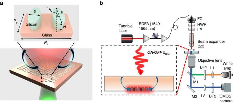
Working principle and experimental facility. a Schematic of the system. When the metasurface is off-resonance, the laser heating of the bulk water induces buoyancy-driven flow, transporting and aggregating particles to the center of the illuminated region. When the quasi-BIC is excited, additional heat sources come from the heat dissipation of the water layer close to the resonators. The thermal-induced flow velocity is increased up to three times. The flow is represented by the two arrows above the nanoantennas. Inset: a unit cell of the metasurface. The geometrical parameters: periods, Px=950nm, Py=778nm; a=532nm, b=192nm, H=190nm, θ=10∘. b Experimental set-up used for excitation of the quasi-BIC metasurface and imaging of the motion of suspended tracer particles. L1 and L2, focusing lenses; M1 and M2, mirrors; BF1 and BF2, bandpass filters used to filter light used for excitation of the fluorescent particles and light transmitted for imaging on the camera, respectively. Filtered fluorescent illumination is passed through the objective lens (10× or 40×) and focused on the sample. EDFA, Erbium-doped fiber amplifier used to amplify the power of the input laser; FC fiber collimator, HWP half wave-plate used to rotate the polarization direction of the laser beam, LP linear polarizer. The metasurfaces and fluorescent tracer particles are visualized on a complementary metal-oxide-semiconductor (CMOS) camera by collecting signals through the same objective lens. Credit: Light: Science & Applications (2023). DOI: 10.1038/s41377-023-01212-4
A cutting-edge practice by two Vanderbilt researchers that enhances light in nanoscale structures could help in the detection of diseases like cancer.
The work by Justus Ndukaife, assistant professor of electrical engineering, and Sen Yang, a recent Ph.D. graduate from Ndukaife's lab in Interdisciplinary Materials Science under Ndukaife, was published in Light: Science & Applications.
In their paper, they show how an engineered nanostructured surface—quasi-BIC dielectric metasurface—can be used to trap micro and sub-micron particles within seconds, which they say helps in the transport of analytes to biosensing surfaces. The metasurface can also serve as a sensor to detect the aggregated particles or molecules, and can be used to enhance fluorescence or Raman signals from the molecules, thereby boosting detection sensitivity, according to the researchers.
"Such a capability could be utilized to detect cancer associated vesicles after aggregating the vesicles for longitudinal patient treatment monitoring and early detection," says Ndukaife, who heads the Laboratory for Innovation in Optofluidics and Nanophotonics (LION) at Vanderbilt.
He adds, "Our work is the first experimental demonstration of the use of quasi-BIC for manipulating fluid flow and suspended particles."
Source: https://tinyurl.com/mr3fxbak via Phys.Org