


 View in
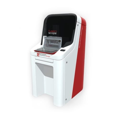
FusionScope
Quantum Design
The FusionScope is a correlative AFM/SEM microscope that has been designed from the ground up to allow you to seamlessly take advantage of combining these two methods of analysis. For most analysis, it is often desirable to analyze a sample with multiple techniques to look for correlations between parameters. For imaging techniques like AFM and SEM, this often means you need to analyze the exact same area. Rather than having to move your sample from one microscope to another, or to use two different operating systems to analyze the same location on a sample, FusionScope provides coordinated complementary measurement on the same location, within the same user interface. This all translates to an easy-to-use system that is capable of quickly providing more useful and novel data.The FusionScope offers all the benefits of a fully functional atomic force microscope with SEM. It is capable of most normal measurement modes expected from a standard AFM, including contact, dynamic, and FIRE modes. Switch between a sub-nanometer resolution AFM and SEM imaging with a simple click of a button to extract your desired data. Interchangeable cantilevers easily provide advanced modes, such as Conductive AFM (C-AFM) and Magnetic Force Microscopy (MFM).
Ease of Use
The FusionScope hardware and software have been carefully designed to allow beginning users to feel comfortable while also allowing advanced users detailed, customizable user interfaces that offer all of the functionality they expect.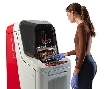
The FusionScope helps users perform common supporting tasks associated with operation of the AFM and SEM. Available selections in the Task Menu allow both new and advanced users to quickly identify and perform the desired microscope operations. The FusionScope software provides a wizard-like experience through its tasks and workflows that guides users through the multi-step processes of operating and configuring the instrument. This experience includes optional animations that helpfully demonstrate the steps involved.
This sophisticated and illustrative task management software helps users spend less time adjusting and managing the hardware and more time gathering sample imagery and making scientific measurements. More advanced users can even coordinate various tasks, ensuring that the chamber is only exposed to atmospheric pressure for a minimum amount of time so the chamber can quickly return to high vacuum to perform SEM imaging. FusionScope Tasks will empower operators to quickly master the operation of an extremely sophisticated scientific instrument.
Sample Exchange
In the FusionScope, changing a sample is fast and easy. The FusionScope software provides helpful task workflows for many routine activities, including sample exchange. These workflows can be accessed through the FusionScope Task menu.
When changing a sample, the FusionScope chamber is first vented to allow the door to be opened. The AFM head is moved to the "park" position so that the sample can be accessed and exchanged without danger of damaging the AFM tip. Once vented the door can be opened. The sample stub must be unscrewed (no screwdriver is needed) and can then be removed and replaced by another loaded sample stub. Close the door and complete the workflow in the software - this will evacuate the chamber again. The complete process takes only a few minutes.
Customization of User Interface
The FusionScope's user-friendly software can be easily adapted to the needs of the user or the experiment. Initially, there are predefined layouts such as a standard mode and an advanced mode. However, depending on which observables are necessary for an experiment, exactly these also can be displayed. A completely individual configuration of the view is possible: position, size and selection of the observables can be set individually. Each "layout" can be saved and called up depending on the user or experiment. Regardless of the choice of observables displayed and the layout, however, all data of all observables are saved permanently and completely - nothing is lost, no matter what is displayed. The lab journal function of the software supports this by allowing metadata and user comments to be recorded in addition to the measurement data. An evaluation of the measurement data is possible both live and offline, as is the export of the data into various other formats.
Data Handling & Post Processing
All data - for both AFM and SEM - acquired for a given sample and AFM tip used are automatically stored in a single "experiment." This makes sure that all related data is kept together in a single project file, which also makes moving data between computers for off-line analysis a convenient and self-organized process. The experiment file stores both the (immutable) original data as acquired, as well as any additional results from post processing (which can be done both during live acquisition as well as after the fact).
Most standard data processing options are available within the desktop application itself and their results can be stored with the experiment. This includes basic corrections (e.g., plane or line corrections for topography data) as well as the ability to perform measurements on an image (the number of individual measurements is not limited).
Additionally, any analysis not currently supported by the desktop application is still possible using third party software, as exporting data to industry-standard file formats (e.g., Gwyddion for AFM data) is built in.
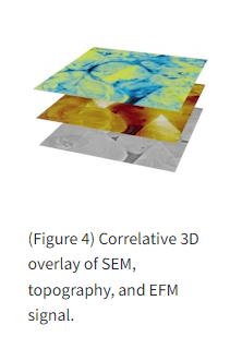
Correlated Coordinate System
At the heart of the FusionScope lies a truly correlated user experience, and key to this is the FusionScope's Correlated Coordinate System. This coordinate system ties together the AFM and SEM operations so that all measurements and tracking are using the same exact "map" to measure your samples and locate specific areas of interest. This also allows for automated features in the FusionScope software, such as locating an area of interest using SEM and then directing the FusionScope to automatically find that same area with the AFM. Gone are the days when a user would have to haphazardly and painstakingly search with the AFM for the particular areas of interest they only just saw using the SEM. Discover the ease-of-use and increased workflow brought to you by the FusionScope Correlated Coordinate System.
Profile View
One of the unique features of the FusionScope is Profile View, where it is possible to observe the tip of the AFM cantilever as it carries out measurements. How is this possible? The AFM scan head and the sample holder are located on a shared trunnion. This allows both parts to be tilted together in relation to the SEM beam. The sample and the tip of the cantilever are simultaneously at the eucentric height of the optics of the electron microscope allowing both to stay in focus when the stage is rotated. This makes it possible to set any tilt angle after the focus has been set on the sample surface and have the selected section on the sample continue to be sharp and visibly in focus. This is nicely demonstrated in the video (below left) which shows a specimen rotating from a tilt angle of 0° to 80° at the eucentric height.
Although the tip length of the AFM cantilever will determine at which angle it will become visible in the electron microscope image, at the maximum tilt of 80° every AFM tip is visible from the side. In addition, the high image acquisition rate makes it possible to observe the movement of the cantilever on the sample surface live. The video (below right) shows this unique capability of the FusionScope.
With Profile View it is also possible to position the AFM tip very precisely on the sample surface. In this way, even hard-to-reach areas of a sample can be approached extremely precisely with the atomic force microscope and complex samples can be measured. In this case, the electron microscope is used to help position the tip. The following images show some impressive examples of this unique capability of the FusionScope.
Self-Sensing Cantilevers
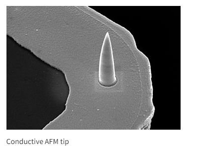
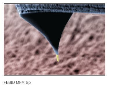
The unique FusionScope Cantilever Technology is based on self-sensing cantilevers that allow for high quality, low-noise detection of surface properties. The cantilevers are equipped with a full piezo-resistive Wheatstone bridge that provides all cantilever signals electrically without the need for optical alignment. This has direct advantages for the performance and ease of use, letting users record better images with less effort. The self-sensing technology also allows maximum access for the electron beam to the cantilever and sample region and enables a seamless combination of AFM and SEM. These self-sensing cantilevers are available with a broad variety of resonance frequencies, spring-constants and tip functionalities which provide the right cantilever for every application. Each cantilever has a unique QR code imprinted on the chip carrier that contains all cantilever-relevant information.
For advanced SPM modes, specialized cantilever tips are used that enable, among other measurements: electrical conductivity, magnetization, surface potentials, temperature, and other sample features. The proper fabrication of such specialized tips is challenging due to the high demands on tip sharpness, long-term mechanical stability, and specific material chemistry for proper functioning. FusionScope offers specialized tips that are fabricated using a unique nano-fabrication method - focused electron beam-induced deposition (FEBID) - that allows for the highest-performance conductive and magnetic tips. The FEBID process enables tip radii of less than 10 nm for high-resolution conductive or magnetic imaging combined with excellent mechanical stability.
The thermal contraction or expansion of mechanical parts within an AFM microscope can sometimes cause unwanted motion in the AFM tip during measurement. This motion is called "thermal drift," and can especially affect measurement of samples smaller than 1um. Identifiable structures on a sample are used to determine the drift rates in the X and Y directions after many scans are taken. Ideally, for good AFMs, drift rates are smaller than 1.5 nm/minute. The FusionScope has been designed to have very low vibrations in the sample chamber by incorporating a floating frame within its structure. This floating frame, along with materials specifically chosen for their stability, allow the FusionScope to experience thermal drift as low as 0.3 nm/min in X and 0.1 nm/min in Y directions.
Applications
By combining the complementary strengths of SEM and AFM, FusionScope opens the door to a whole world of new application possibilities.Use the FusionScope for detailed Material Characterization of your samples and perform correlative analysis of their structural, mechanical, electrical, magnetic, and chemical properties on exactly the region of interest.
Whether you are looking for high-level Quality Control of component parts or want to perform Failure Analysis on electrical components or semiconductor devices, FusionScope will help you to get the job done. Benefit from the fast and intuitive workflow to extract the data you are looking for.
Combining high-resolution SEM and state-of-the-art AFM you can easily characterize Nanostructures such as nanowires, 2D-materials, and nanoparticles. FusionScope gives you full control to locate the Nanostructures and perform the measurements of your choice.
Using FusionScope in Life Science applications allows you to acquire the nanoscale morphology of biological samples accurately and easily. Especially for hard-to-reach sample areas or very small features, FusionScope allows you to characterize physical properties such as 3D topography, stiffness, and adhesion with the highest precision.
Materials Characterization
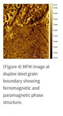
Characterize Magnetic Phase Structures using Magnetic Force Microscopy
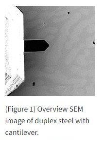
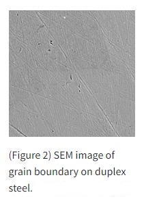
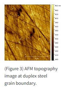
Duplex is a family of stainless steel grades that contain a mixture of austenitic and ferritic phases that provide higher mechanical strength and ductility compared to standard steel grades. In-situ Magnetic Force Microscopy (MFM) enables the detailed analysis of the magnetic properties of different types of duplex steel samples.
With the FusionScope the different phases of the steel surfaces can be visualized, and the cantilever is easily positioned at the grain boundary of two distinct phases. Using a magnetic cantilever tip the magnetic properties can be analyzed, and the ferromagnetic sub domains can be imaged with high resolution.
 Characterize Magnetic Phase Structures using Magnetic Force Microscopy
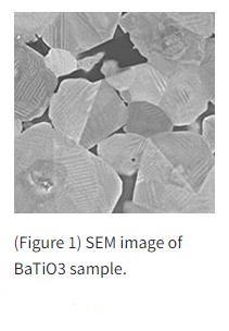
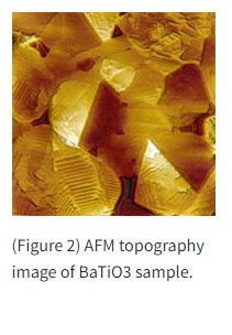
Barium titanate (BaTiO3) is a ceramic material exhibiting interesting optical, electrical, and thermal properties shifting it to the center of scientific attention. More recently BaTiO3 is gaining importance also for engineering applications. Ferroelectric BaTiO3 is a non-linear positive-temperature-coefficient (PTC) material and is used in resistors. Polycrystalline doped barium titanate exhibits a wide range of electrical resistance depending on the temperature which is employed in sensors and actuators.
The macroscopic electronic properties of polycrystalline BaTiO3 ceramics are governed by potential barriers forming between single grains. To reach a better understanding of the overall resistance of barium titanate it is essential to be able to characterize the potential differences in the crystalline material at the nanoscale.
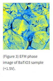
This characterization can be done with Electrostatic Force Microscopy (EFM). It is widely used in electronics development to map electronic characteristics of complex, sub-micron electrical materials. FusionScope enables the possibility for in-situ EFM analysis. The high resolution of the SEM can be used to easily identify grain boundaries and perform the EFM analysis directly at the region of interest.
 Quality Control & Failure Analysis
Analyze Materials with Difficult Geometries using Atomic Force Microscopy
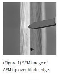
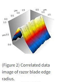
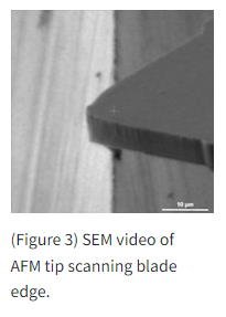
Typically, in atomic force microscopy, measurements of very pointed sample geometries are difficult. Firstly, due to the convolution of the geometry of the tip with the topography of the sample surface, but also the correct and reliable positioning of the tip over the sample is a challenge. SEM is used here to position the sample in the best possible way and to monitor the AFM measurement in real time.
A commercially available razor blade was installed in the sample holder with the aim of imaging the surface of the blade with the AFM and, in particular, determining the radius of the blade edge. The measurement comprises several steps: the coarse positioning, the fine positioning, the approach of the tip and finally the measurement of the topography. With the help of the fine positioning made possible by the SEM, different areas on the razor blade can be quickly selected and measured. Different material properties, such as a coatings applied to the razor blade, also can be compared. An important parameter is the radius of the razor blade, as well as the roughness of the surface.
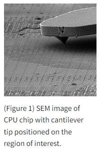
Analyze Electronic Components or Semiconductor Devices using Atomic Force Microscopy
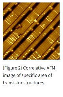
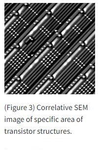
Detailed location and analysis of nanometer-sized structures is a challenging and time-consuming task for all AFM operators. The size reduction in recent generations of transistors creates especially high demands on quality control and failure analysis. With FusionScope and Profile View you can easily navigate the cantilever tip to the region of interest and perform high resolution AFM analysis of your sample. Measure the real 3D topography with sub-nanometer resolution or extract additional information using conductive AFM.
Specifications
AFM |
Scan Range XY: |
22 x 22 µm (Closed Loop) |
Scan Range Z: | 15 µm | |
Imaging Noise: | <50 pm @ 1 kHz | |
Cantilever Probes: | Self-Sensing Piezoresistive | |
Measurement Modes: | Contact, Dynamic, FIRE, MFM, C-AFM, | |
SEM |
Electron Source: |
Thermal Field Emission |
Acceleration Voltage: | 3.5 kV 15 kV | |
Probe Current: | 5 pA 2.5 nA (300 pA typical) | |
Magnification: | 25X 200,000X | |
Detectors: | In-Chamber SE Everhart-Thornley | |
SAMPLE |
Max Sample Diameter: |
20 mm (12 mm Full Correlated Mode) |
Max Sample Height: | 20 mm | |
Max Sample Weight: | 500 g | |
Eucentric Alignment: | Automatic | |
CHAMBER |
Typical Chamber Vacuum: |
1-10 µTorr |
Pumping Time: | <5 min | |
Trunnion Tilt: | -10 to 80 Degrees | |
SYSTEM |
Power: |
200-230 VAC; 50/60 Hz; Single Phase 15 A |
Dimensions (W x L x H): | 690 x 835 x 1470 mm | |
Weight: | 330 kg |
Videos


